Price List Knee Surgery
| Knee Surgery and Replacement | Average Hospitalization | Average Total Cost* |
| Complex Knee Arthroscopy / Meniscus Repair (Minimally Invasive Knee Surgery) | 2 nights | 10.000 € |
| Osteotomy for Knee Correction | 3 nights | 15.500 € |
| Surgery for Patella (Knee Cap) Dislocation | 2 nights | 13.000 € |
| Partial Knee Prosthesis (Unicompartmental Knee / Repicci) | 5 nights | 21.500 € |
| Total Knee Replacement | 5 nights | 23.300 € |
| Total Knee Replacement Revision Surgery | 7 nights | 36.500 € |
| Knee Cartilage Transplant (ACT), harvesting, cultivation and transplant | 2+3 nights (2 procedures) | 31.200 € |
| Anterior Cruciate Ligaments Reconstruction | 2 nights | 13.000 € |


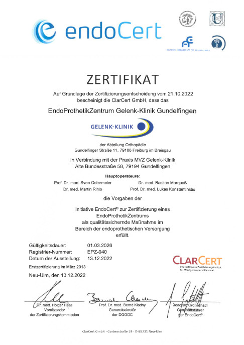


 certifies the superior quality of patient consultations, diagnostics, surgery and after-care for foot and ankle conditions and injuries at the orthopedic hospital Gelenk-Klinik, Gundelfingen.")
 or impact on the knee. When it is ruptured, we refer to a cruciate ligament tear. © Istockphoto.com/MedicalArtInc")


 ©Viewmedica")