Malunion ankle fracture: early treatment prevents arthrosis
- Long-term consequences of a malunion ankle fracture: arthrosis
- What are the symptoms of a malunion ankle fracture?
- Treatment options for malunion ankle fractures
- Orthopaedic surgeon and foot and ankle specialist for ankle surgery at Gelenk-Klinik
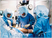
- Anaesthesia for ankle surgery
- Aftercare, rehabilitation following ankle surgery
- Should I expect pain after ankle surgery?
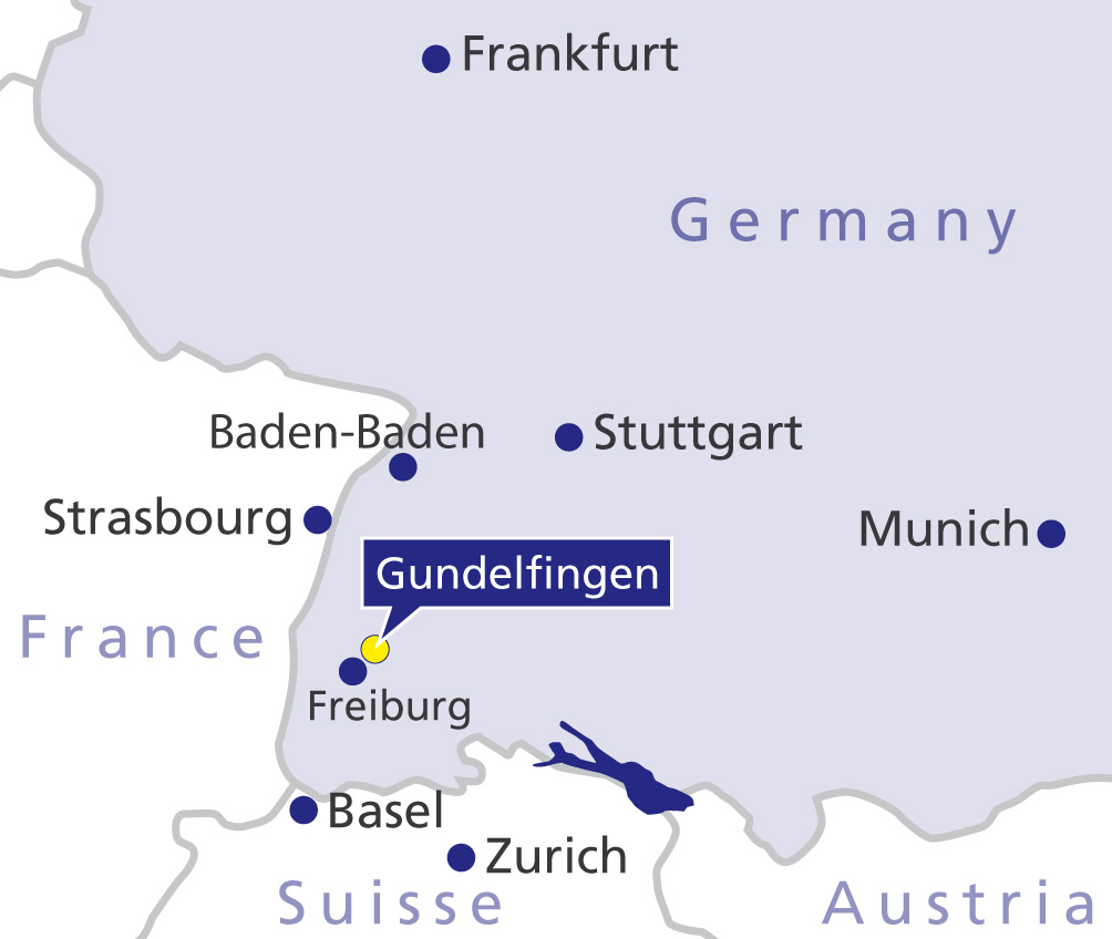
- How will my treatment be organized at Gelenk-Klinik?
- Important considerations after surgery
- Cost of ankle surgery in Germany
- Scheduling your appointment for ankle surgery in Germany
 is only bearing weight at the lateral malleolus (right) due to the angulation. The unilateral load can cause arthrosis in patients. © Gelenk-Klinik") X-ray of a malunion ankle fracture. The joint surfaces in the ankle are not lined up properly and are no longer parallel. The talus (bottom) is only bearing weight at the lateral malleolus (right) due to the angulation. The unilateral load can cause arthrosis in patients. © Gelenk-Klinik
X-ray of a malunion ankle fracture. The joint surfaces in the ankle are not lined up properly and are no longer parallel. The talus (bottom) is only bearing weight at the lateral malleolus (right) due to the angulation. The unilateral load can cause arthrosis in patients. © Gelenk-Klinik
The term malunion ankle fracture refers to a fracture in the bones maintaining the joint that did not heal correctly. This can occur after rolling and twisting, e.g. during sports but also after falling.
If the fracture does not heal correctly, deformities or different levels can form on the joint surface in the ankle. Patients are afflicted with chronic pain and limited mobility. Abnormal biomechanical stress of the joint causes cartilage damage in the long term with subsequent ankle arthrosis.
At Gelenk-Klinik, experienced foot and ankle specialists competently examine and treat malunion ankle fractures and remaining dysfunctions. The proposed treatments are tailored to the patient's specific situation, e.g. the age or favourite sports.
Long-term consequences of a malunion ankle fracture: arthrosis
Ankle fractures can occur in various areas of the bones. The fracture is often accompanied by injuries to the ligament complex - primarily the lateral ligaments.
If the bone fragments in the ankle are not positioned exactly parallel as the ankle is healing, deformities and changes in the shape of the joint can occur. These malunions of the ankle are very painful and could have serious consequences:
- limited mobility and blockages in the ankle
- painful weight-bearing, limiting physical activity
- permanent instability of the ankle along with frequent twists
- chronic ankle pain
- early cartilage wear (arthrosis) due to the deformity and change in the shape of the surfaces on the inside of the ankle
Which types of malunion ankle fractures are there?
Types of malunion ankle fractures:
- pseudarthrosis (false joint)
- axial or rotation deformities of the bone
- bone deformities
- shortened or lengthened bones
- ligament instabilities (lateral ligament and syndesmosis ligament)
Ankle fractures are fractures in the ankle area and are classified as different types of fractures:
- isolated fractures of the lateral malleolus: calf bone (fibula)
- isolated fractures of the internal malleolus: shin bone (tibia)
- combined fracture of both parts of the ankle: bimalleolar ankle fracture
Ankle fracture classification according to Weber
 Lateral malleolus fractures are distinguished according to the Weber classification. The classification depends on how high on the lower fibula the fracture is and whether the syndesmosis ligament is involved. © Gelenk-Klinik
- Weber-A: The fracture is below the level of the syndesmosis (ligament connecting the tibia and fibula), and the syndesmosis is intact.
- Weber-B: With a type Weber-B injury, the area around the syndesmosis is affected by the fracture. The syndesmosis is often also injured.
- Weber-C: With this type, the fracture is above the level of the syndesmosis. The syndesmosis is also injured. The stabilizing membrane between the tibia and fibula (interosseous membrane of the leg) is often also injured.
Even with conservative treatment and surgery for a lateral ankle fracture, so-called pseudarthrosis can develop. The term pseudarthrosis comes from Greek (pseudos = false, arthros = joint) and refers to a false joint forming due to a bone malunion. The affected fibula does not heal properly following a fracture.
The internal malleolus fracture can also result in bone deformities, bone shortening or even lengthening of the tibia, thus joint damage.
Fractures in the area of the posterior joint surface of the tibia, the so-called Volkmann's triangle, can cause step-offs or gaps to form on the joint surface. Step-offs lead to severe ankle arthrosis in patients in the long term. Due to accompanying tears to the medial ligaments, the supporting ligament complex becomes unstable and the joint bears weight incorrectly in the long term.
Special type of ankle fracture: the Maisonneuve fracture
The Maisonneuve fracture is a special type of ankle fracture. In addition to an internal malleolus fracture or medial ligament rupture, there is a fibula fracture near the ankle. The syndesmosis is also torn in Maisonneuve fractures, and often also the stabilizing membrane between the two lower leg bones (interosseous membrane of the leg). A possible long-term consequence is a deformity in the talar mortise.
What are the symptoms of a malunion ankle fracture?
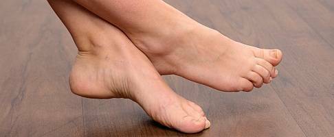
The subsequent damage of a malunion ankle fracture can start early, within months, but sometimes even after decades. Patients with malunion ankle fractures particularly report noticeable limitations in the ankle. They experience reduced mobility and foot roll-over is not optimal when walking.
Six months after a fracture, the patient should be able to move and use the ankle in daily activities and sports without pain.
There are typical symptoms which can occur in a malunion ankle fracture:
- swollen ankle,
- limited foot roll-over,
- the ankle locks while straightening or bending,
- weight-bearing on the ankle is painful, patients are unable to walk as far as they used to,
- the ankle is permanently unstable, the patient frequently twists their ankle.
- the patient has chronic ankle pain,
- the patient is experiencing discomfort and limitations in the ankle following a fracture, lasting more than 6 months,
- the area around the fracture is infected.
Treatment options for malunion ankle fractures
The goal of medical treatment for a malunion ankle fracture is always to restore the natural shape of the joint so the joint can fulfil its function optimally. The outcome of treatment depends on the following factors:
- extent of the injury
- accompanying cartilage damage on the joint surface
- soft tissue injuries and scar tissue formation
- patient's age and gender
- history of conservative or surgical initial care following the ankle fracture
 A detailed physical examination serves as the basis for further treatment. © joint-surgeon
Osteotomy: surgical realignment of the ankle bones
Up to what stage joint-preserving surgery is performed is determined by the specialist based on the patient's needs. The specialist and patient discuss joint-preserving treatment options based on the damage to the ankle.
The joint cannot always be restored to its original condition with realignment surgery. Even if surgery is successful, there may still be limitations in ankle mobility or sensitivity disorder in the foot.
However, a surgical procedure will yield considerable long-term improvement in symptoms, even in patients suffering greatly due to advanced wear in the ankle. In these cases, the foot and ankle specialist will correct the cause of arthrosis by correcting the position of the bones in the ankle (osteotomy). The orthopaedist performs the osteotomy on the lateral malleolus, internal malleolus or on the joint surface of the tibia, depending on where the fracture is located.
By refixating the malaligned bones and then healing in the new position, the foot and ankle specialist can often considerably improve the anatomical shape and thus increase the weight-bearing capacity of the ankle.
 reduces the fit of the other joint parts in the ankle, most of the weight is on the inside (medial) edge of the anklebone. Arthrosis develops here due to chronic strain. Right: After surgical repositioning of the bone (osteotomy) of the shinbone (tibia) the entire joint surface is fully weight-bearing again. The osteotomy can – depending on the initial situation – stop ankle arthrosis or delay it by many years. © Gelenk-Klinik") Left: The tilt in the anklebone (talus) reduces the fit of the other joint parts in the ankle, most of the weight is on the inside (medial) edge of the anklebone. Arthrosis develops here due to chronic strain. Right: After surgical repositioning of the bone (osteotomy) of the shinbone (tibia) the entire joint surface is fully weight-bearing again. The osteotomy can – depending on the initial situation – stop ankle arthrosis or delay it by many years. © Gelenk-Klinik
Left: The tilt in the anklebone (talus) reduces the fit of the other joint parts in the ankle, most of the weight is on the inside (medial) edge of the anklebone. Arthrosis develops here due to chronic strain. Right: After surgical repositioning of the bone (osteotomy) of the shinbone (tibia) the entire joint surface is fully weight-bearing again. The osteotomy can – depending on the initial situation – stop ankle arthrosis or delay it by many years. © Gelenk-Klinik
During surgery on a malunion ankle fracture, the ankle specialist assesses the entire ligament complex. Injuries to the syndesmosis, medial and lateral ligaments are corrected immediately by ligament reconstruction if required.
Regeneration of the joint surface through autologous cartilage transplantation (ACT)
The orthopaedic ankle specialist can correct localized cartilage damage in the malunion ankle with the autologous (body's own) cartilage transplantation (ACT). As part of the joint-preserving surgery, the surgeon harvests a tiny amount of cartilage cells from an intact area in the patient´s joint. The cartilage cells are grown in the laboratory. The resulting mass of cartilage cells is reimplanted in order to reinforce damaged cartilage area after about 6–8 weeks.
Ankle prosthesis: artificial joint replacement
The goal of an ankle prosthesis is to restore pain-free and natural movement in the ankle for advanced arthrosis due to the malunion fracture. The particular advantage of the ankle prosthesis is preserving the normal gait.
A prosthesis requires that the joint is correctly aligned. Damage to the ligament complex or bone deformities in the ankle must be corrected beforehand. Thus joint-preserving ankle surgery (osteotomy) can precede a prosthesis implantation. Correcting the malalignment of the ankle sometimes considerably reduces pain and the planned joint replacement can be postponed because it is no longer necessary.
If the prerequisites for an ankle prosthesis are not met, the specialists at Gelenk-Klinik will suggest fusing the ankle joint.
Ankle arthrodesis: ankle fusion surgery
An arthrodesis is an artificially created bone union between the lower leg bones and the foot. During a fusion of the talocrural joint, the talus is fixed to the ankle mortise above it with screws. The bones in the joint grow together permanently as a result of the arthrodesis. Mobility in the joint is completely eliminated in favour of a stable bone connection between the joint parts. The fusion allows the patient to enjoy pain-free full weight-bearing on the ankle previously afflicted with arthrosis. Arthrodesis also eliminates any pre-existing joint instability, compensating for any instability of the ligaments.
However, ankle fusion surgery always involves a substantial limitation in ankle mobility. The ankle arthrodesis noticeably changes the natural gait pattern. The altered motion sequence has the potential to strain nearby joints of the foot and - while rendering the ankle pain-free and with maximum weight-bearing capacity - slightly increases the possibility of developing arthrosis in the hip, foot or talotarsal joint.
Orthopaedic surgeon and foot and ankle specialist for ankle surgery at Gelenk-Klinik
Here at Gelenk-Klinik, a close relationship between the physician and their patient is important. This means: You will be in the care of your treating physician from the initial examination until after surgery. So, you will always have one contact person who will be very familiar with your case and assigned to you throughout your stay at Gelenk-Klinik. Our experts in foot and ankle surgery are experienced specialists Dr. Thomas Schneider and Dr. Martin Rinio. They are certified foot and ankle surgeons whose continuing education, diagnostics and surgical quality is reviewed annually by an independent German professional society (DAF). The orthopaedic Gelenk-Klinik is therefore a speciality centre with the title "Centre for foot and ankle surgery" (ZFS).
Anaesthesia for ankle surgery
We typically perform ankle surgery under general anaesthesia. However, we also offer spinal anaesthesia to avoid general anaesthesia if the patient so desires. In the case of spinal anaesthesia, an anaesthetist injects the anaesthetic into the spinal canal of the lumbar spine. The patient is fully conscious during surgery. You will decide which type of anaesthesia is best for you together with the anaesthetist. The anaesthetists at Gelenk-Klinik are very experienced in both methods and will choose the option best for the patient and their circumstances based on a pre-operation discussion.
Aftercare, rehabilitation following ankle surgery
After ankle surgery, you are only allowed partial weight bearing and will therefore have forearm crutches. Your foot will be stabilized in a special Orthosis, which you will be wearing day and night. We will ensure you receive the aid required after surgery.
Thrombosis prophylaxis (e.g. using Heparin/Enoxaparin) is therefore vital. This will prevent dangerous blood clots from forming. We strongly recommend physiotherapy and lymphatic drainage after leaving the hospital to prevent muscle loss and minimise swelling of the forefoot. The older the patient, the longer the forefoot will be swollen.
Should I expect pain after ankle surgery?
Every procedure causes pain. We always strive to keep pain after ankle surgery as low as possible. The assigned anaesthetist will already start pain management during the procedure. In many cases the anaesthetist uses a so-called nerve block during surgery, which keeps the respective foot pain free for approx. 30 hours. This already manages the greatest wave of pain, which can then be easily treated with regular medication. Our goal is for you to experience as little pain as possible after surgery.
How will my treatment be organized at Gelenk-Klinik?
 Private room in the Gelenk-Klinik in Gundelfingen, Germany. © joint-surgeon
During your inpatient stay at Gelenk-Klinik you will have a single-occupancy room. The room has a bathroom with a shower and WC. All rooms include towels, a bathrobe and slippers. They further include a mini-bar and a safe. All rooms also have a television. You only need to bring your personal medication, comfortable clothes and sleepwear. After surgery, you will receive 24-hour care from experienced nursing staff and experienced physiotherapists. The inpatient stay following the procedure is typically 3 days. Your family members can stay at a hotel within walking distance. We will gladly take care of making the reservations.
Important considerations after surgery
Depending on the procedure performed, there are different situations you will have to have in mind as a patient after the surgery.
Osteotomy:
- Weight bearing: partial weight bearing of 20 Kg for 12 weeks
- VACOped Orthosis for 12 weeks
- Inpatient treatment: 5 days
- Optimal local stay: 12 days
- Earliest return flight: 10 days after surgery
- Recommended return flight: 15 days after surgery
- Showering permitted: 12 days after surgery
- Recommended time off work: 6 -12 weeks (depending on the work)
- Recommended removal of sutures: 12 days
- Time before being able to drive again: 4 month
- Rehabilitation: can be started after 12 weeks
- Sports: soonest after one year
Prosthesis:
- Weight bearing: partial weight bearing of 20 Kg until wound healing is complete, then slow increase of weight: half body weight at the end of week 6
- VACOped Orthosis for 6 weeks
- Inpatient treatment: 5 days
- Optimal local stay: 12 days
- Earliest return flight: 10 days after surgery
- Recommended return flight: 15 days after surgery
- Showering permitted: 12 days after surgery
- Recommended time off work: 6 weeks (depending on the work)
- Recommended removal of sutures: 12 days
- Time before being able to drive again: 8 weeks
- Rehabilitation: can be started after 8 weeks
- Sports: soonest after one year
Arthrodesis:
- Weight bearing: partial weight bearing of 20 Kg for 8 weeks - X-ray control at this stage decides about the following increase in weight bearing
- VACOped Orthosis for 8 weeks
- Inpatient treatment: 5 days
- Optimal local stay: 12 days
- Earliest return flight: 10 days after surgery
- Recommended return flight: 15 days after surgery
- Showering permitted: 12 days after surgery
- Recommended time off work: 8 weeks (depending on the work)
- Recommended removal of sutures: 12 days
- Time before being able to drive again: 10 weeks or more, depending on the X-rays
- Rehabilitation: can be started after 12 weeks
- Sports: soonest after one year
Cost of ankle surgery in Germany
In addition to the cost of surgery, you will also need to plan for additional costs for diagnostics, physician’s appointments and aids (e.g. forearm crutches). These will be about 1,500 to 2,000 Euros. If you plan to have outpatient physiotherapy after surgery, we will gladly provide an estimate of costs. You will find information about the cost of hotel lodging and any follow-up treatment at the rehabilitation clinic on the websites of the respective providers.
Scheduling your appointment for ankle surgery in Germany
The foot specialists in Germany will first need current MRI images and x-rays of the respective foot. After sending these documents to us through our website, we will send you patient information including proposed treatment and a binding estimate of costs within 1–2 days.
Gelenk-Klinik offers quick appointments for international patients to correspond with your preferred travel itinerary. We will gladly help you apply for a visa once we have received the down payment specified in the cost estimate. If the visa is denied, we will promptly refund the full down payment by remittance.
We try to minimise the time between the preliminary examination and the surgery for our international patients to avoid additional travel. During your inpatient and outpatient stay our multilingual (English, Russian, Spanish, Portuguese) case management team will be there to assist you. We can also provide an interpreter (e.g. Arabic) at any time, at the patient’s expense. We will gladly assist you with organising transportation and lodging and provide tips for recreational activities for your family members.